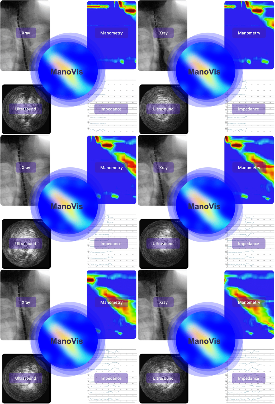
Patients with benign diseases of the esophagus often have a long medical history until their final diagnosis. Depending on severity and symptoms of the disease, patients undergo a wide variety of examinations such as EGD, manometry, pH-metry, CT/MRI, videofluoroscopy, FEES, etc. While all examination results are compiled piece by piece and gradually depict the entire aspects of the disease, it is still extremely difficult to link the individual examination modalities with each other.
The aim of our project is to develop a combined diagnostic means that allows optimal visualization of esophageal motility. The combined analysis method should not only lead to more intuitive findings, but rather to a reduction in examination time for both patient and physician, as well as to a reduction in exposure to ionizing radiation in the long term.

