Laboratories
Our research innovates hybrid methods combining thermoacoustic, optoacoustic and fluorescence imaging. Fueled by a DFG Koselleck award, and BMBF support (Tech2See, Sense4Life) award we innovate in three main areas; (1) We interrogate the fundamental interactions of electromagnetic (EM) and optical energy and tissue and exploit them for sensing applications. (2) We research novel instrumentation designs for optimal illumination and energy coupling to tissue for sensing and therapeutic applications and (3) develop novel classes of sensors (including nanoparticles and novel sensing devices) for reading pathophysiological features of tissue, indicative of disease aiming to generate novel early detection paradigms. System development, data processing and in-vivo monitoring of biophysical processes are among the focus of our research group. Research from this group further feeds technological solutions in other research groups in the Chair and Institute.
Relevant publications
Omar M, Soliman D, Gateau J, Ntziachristos V. Ultrawideband reflection-mode optoacoustic mesoscopy, Optics Letters 39(13); 3911-3914 (2014)
George J. Tserevelakis, Dominik Soliman, et al. Hybrid multiphoton and optoacoustic microscope. Optics letters 39.7 (2014): 1819-1822. Omar M, Gateau J, Ntziachristos V. Raster-scan optoacoustic mesoscopy in the 25-125 MHz range, Optics Letters 38(14); 2472-2474 (2013)
Kellnberger S, Deliolanis NC, Queirós D, Sergiadis G, Ntziachristos V. In vivo frequency domain optoacoustic tomography, Optics Letters 37(16); 3423-3425 (2012)
Omar M, Kellnberger S, Sergiadis G, Razansky D, Ntziachristos V. Near-field thermoacoustic imaging with transmission line pulsers, Med. Phys. 39(7); 4460-6 (2012)
Kellnberger S, Hajiaboli A, Razansky D, Ntziachristos V. Near-field thermoacoustic tomography of small animals, Phys. Med. Biol. 56(11); 3433-44 (2011)
Razansky D, Kellnberger S, Ntziachristos V. Near-field radiofrequency thermoacoustic tomography with impulse excitation, Med. Phys. 37(9); 4602-4607 (2010)
Research Highlights
Thermoacoustic imaging of small animals at high spatial resolution
The thermoacoustic effect describes the dissipation of transient electromagnetic (EM) energy in tis-sue followed by the induction of an acoustic wave owing to thermoelastic expansion. In thermo-acoustic imaging, objects are irradiated with short EM pulses of high energy to induce acoustic waves, yielding an imaging contrast which is based on the electric and dielectric properties of tissue. To deposit sufficient energy in tissue, conventional thermoacoustic tomography (TAT) approaches prolong the EM pulse duration at the cost of spatial resolution, limiting TAT from resolving submilli-metre structrures. To overcome resolution and energy coupling limitations, we propose near-field coupling of the object to the energy emitting element (antenna) in combination with ultrashort RF pulses. Our near field radiofrequency thermoacoustic (NRT) tomography approach relates to stimula-tion of biological tissue with nanosecond high energy pulses instead of carrier frequency amplifica-tion employed in conventional TAT. We designed dedicated high voltage impulse generators, provid-ing < 100 ns pulses carrying energies of hundreds of mJ. Employing transmission line pulsers, we could reduce the impulse duration without compromising the pulse energy, enabling non-invasive small animal imaging at high spatial resolution (see Omar M, 2012 Med Phys. 39(7); 4460-6)).
Raster-Scan Optoacoustic Mesoscopy
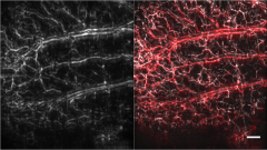
In raster-scan optoacoustic mesoscopy (RSOM) we focus on imaging the mesoscopic gap, which re-fers to the depths beyond what microscopic techniques can image, i.e. deeper than 500 µm, and before macroscopic techniques become efficient in imaging, i.e. up to 5 mm in depth. The range of applications at this depth include imaging of model organisms used in developmental biology, skin imaging, and imaging the microenvironment of diseases.
Hybrid optical and Optoacoustic Microscopy
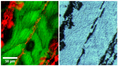
The combination of optoacoustic imaging with optical microscopy techniques offers several ad-vantages. First, the use of label-free modalities allows for imaging of various disease biomarkers without interfering with biological systems. Second, the integration of RSOM enables multi-scale imaging, where optical microscopy techniques allow for high resolution imaging of smaller regions at the first few hundreds of microns, while optoacoustic mesoscopy facilitates whole-organism imaging at larger depths of several millimeters. Finally, the combination of optical and optoacoustic imaging techniques provides visualization of different anatomical markers with complimentary contrast, ena-bling broader information to be obtained from complex biological organisms. Examples of optical microscopy modalities that are combined with optoacoustic imaging are selective plane illumination microscopy (SPIM), as well as non-linear microscopy techniques such as two-photon excitation fluo-rescence imaging (TPEF), second harmonic, and third harmonic generation microscopy (SHG / THG).

