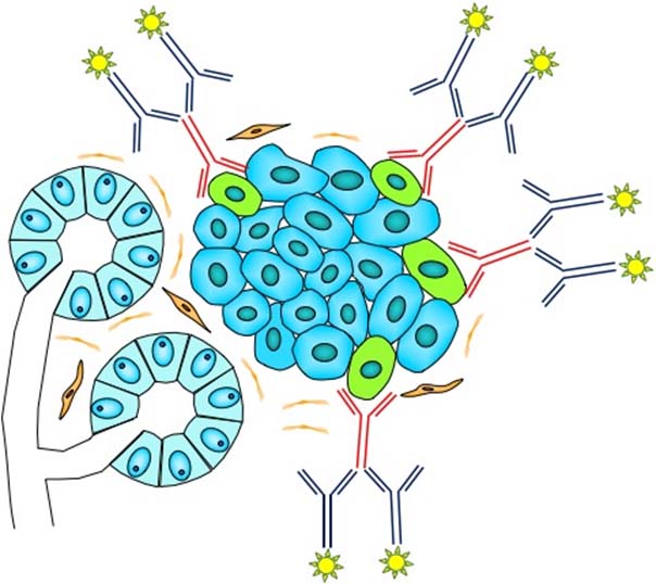
Immunohistochemistry is widely used in modern pathology to identify and visualize proteins (in this context called antigens) within tissue sections (and cell blocks). For better sensitivity and specificity, we are using the indirect approach wherein a subsection of the antigen (a so called epitope) is targeted with a specific primary antibody. Visualization is achieved using a secondary antibody coupled to an enzymatic reporter complex that will bind specifically to the primary antibody. The enzymatic reactions results in precipitation of a brown or red colored dye within the cellular compartment where the antigen is located. In order to visualize the morphology of the tissue, each slide is additionally stained with hemalum. In general, primary antibodies are derived from mouse or rabbit and secondary antibodies are directed against the respective species.
Since results of immunohistochemistry are influenced by a number of factors (e.g. tissue fixation) protocols are optimized, standardized and each stained is accompanied by on slide controls. Additionally, our protocols are validated by participating in certified ring trails (e.g. QUiP). The complementary use of immunohistochemistry together with traditional stains (e.g. hematoxylin and eosin) can aid in the diagnostic process, especially in delineating different tumor subtypes. A large number of different antibodies and combinations thereof can for example enable the identification of a primary tumor in patients presenting with metastases. Even tumors with similar morphology can be distinguished from one another using immunohistochemistry. This is based on the fact that different cell and tissue types are characterized by different (glyco-)proteins located within nucleus, cytoplasm or cell membrane. These include cytokeratins, intermediate filaments, hormones, enzymes, cell cycle proteins, and membrane proteins. Other fields of application include detection of viral proteins (e.g. EBV or CMV), identification of drugable targets (e.g. Her2, PD-L1, NTRK, SSTR2a), and tumor differentiation (MIB1 index). The lab is equipped with three strainers in order to perform roughly 200 immunohistochemical stains per day. For routine diagnostics, more than 220 different antibodies are included in our repertoire.