Figure: CBI
The Clinical Bioengineering Group researches (multi-spectral) optoacoustic imaging technology geared towards clinical applications. Multi-Spectral Optoacoustic Tomography (MSOT) is a novel and powerful imaging modality pioneered at IBMI which offers high-resolution optical imaging through several millimeters to centimeters of tissue. While optical imaging is typically limited by photon scattering, resulting in image blurring, the unprecedented performance of MSOT breaks through the traditional barriers of optical imaging and microscopy (e.g. Ntziachristos V. et al., Nature Methods 7(8), 2010) and has already enabled novel insights into functional, molecular and dynamic parameters in biological and medical imaging (e.g. Herzog E. et.al., Radiology 263(2), 2012). To enable cutting edge research within the imaging sciences the laboratory has strong expertise in following areas:
Real-time and handheld imaging technology for optoacoustic tomography, including hardware design and prototype system development. This activity has now developed several imaging systems for cross-sectional real-time macroscopic tissue imaging, most notably the MSOT II small animal imaging system (e.g. Buehler et. al., OPTICS LETTERS 35(14), 2010), 3D imaging systems (e.g. Buehler et al. Optics Express 20(20); 2012) and handheld systems for clinical use (e.g. Buehler et al. , Optics Letters, 38(9), 2013) which resulted, among other results, in the first real-time optoacoustic images, first real-time multispectral images or the first ever produced images of the human carotids (e.g. Dima et al., Optics Express, 20(22), 2012) and thyroid (Dima et al., Photoacoustics, 4 (4), 2016).
Novel Tomographic Theory including forward and inversion methods geared to optimally utilize the rich, wide-bandwidth information data set collected by MSOT systems to generate accurate optoacoustic images. Model-based mathematical approaches have been developed for image reconstructions of broadband data and implementation of detector impulse response (e.g. Queiros et. al. Journal of biomedical optics 18(7), 2013).
Spectral unmixing methods for physiological and molecular imaging allowing non-invasive probing of tissue physiological states, resolving pharmacokinetics and providing molecular images, including imaging of various extrinsically administered agents such as circulating fluorescent dyes, targeted fluorescence probes and gold or carbon nanoparticles or visualization of physiological parameters such as tumor hypoxia (e.g. Tzoumas et al. IEEE, TMI-2016-0326).
Endoscopic optoacoustic methods for anatomical and molecular imaging and non-invasive probing of tissue physiological states inside the gastrointestinal tract and other luminal organs.
Relevant publications
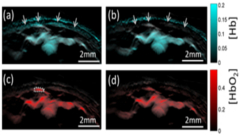
Taruttis A., Timmermans A.C., Wouters P.C.,Kacprowicz M., Gooitzen M van Dam, Ntziachristos V. Optoacoustic Imaging of Human Vasculature: Feasibility by Using a Handheld Probe Radiology, Vol 281, Issue 1, 2016
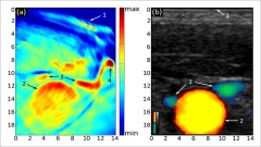
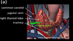
Dima A., Ntziachristos V. In-vivo handheld optoacoustic tomography of the human thyroid Photoacoustics, Volume 4, issue 2, 2016
Buehler A., Kacprowicz M., Taruttis A., Ntziachristos V. Real-time handheld multispectral optoacoustic imaging Optics Letters, Vol. 38, Issue 9, pp. 1404-1406 (2013)
Dima A., Ntziachristos V. Non-invasive carotid imaging using optoacoustic tomography Optics Express, Vol. 20, Issue 22, pp. 25044-25057 (2012)
Razansky D, Buehler A, Ntziachristos V. Volumetric real-time multispectral optoacoustic tomography of biomarkers Nat Protoc, 6(8);1121-9 (2011)
Research Highlights
MSOT is a five-dimensional multi-scale imaging approach, when considering the 3 geometrical dimensions, the spectral dimension (imparting physiological and molecular imaging) the time-dimension (up to 100 frames per second) which enables imaging of dynamic biological phenomena and pharmacokinetics. These developments are geared to advancing basic research, drug discovery and clinical decision making. Work in the group has resulted in a technology platform that has attracted several awards and grants including a senior ERC grant, a European FP7-ICT grant (FAMOS), the GO-Bio Innovation award and a Medical technology innovation award from the Federal Ministry of Education and Research (BMBF) in Germany.
Real-time MSOT

Figure: CBI
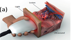
Multispectral optoacoustic tomography (MSOT) is an emerging non-invasive bio-medical imaging technique based on the optoacoustic effect, i.e. the generation of ultrasound after absorption of light energy. MSOT can resolve optical absorption contrast deep inside living biological tissue in real-time with unprecedented high resolution and due to the use of multiple wavelengths with molecular specificity. These distinct properties of MSOT offer major opportunities for visualizing basic biological processes at an anatomical, physiological and molecular level and thus improving clinical diagnosis and treatment of cancer and other major diseases (see Buehler A. et. al., 2013 Optics Letters, 38(9), Razansky D. et al. 2011 Nat Protoc, 6(8), Buehler A. et al. 2010 OPTICS LETTERS 35(14)).
Non-invasive clinical optoacoustic imaging

Figure: CBI

Figure: CBI
The combination of optical contrast and ultrasound resolution at tissue depths beyond a few millimeters renders optoacoustic imaging a promising tool for clinical diagnostic imaging. Stationary tomographic systems for breast cancer detection and handheld systems based on linear ultrasound arrays have already been investigated, but had no significant impact due to limited sensitivity, tissue depth, image quality or a combination of all three. To unleash the true potential of optoacoustic imaging we are thus developing a new generation of non-invasive handheld systems that are better adapted to the optoacoustic imaging paradigm of tomographic detection. We have engineered an imaging system for imaging 2-3cm below the skin surface with resolutions of up to 150µm at up to 50 image frames per second (see Dima A. et al., 2011 Expert Opinion on Medical Diagnostics 5, 263-272, Dima A. et al., 2012 Opt. Express 20, 25044-25057, Buehler A. et al., 2013, Optics Letters, 38(9), Diot G. et al. 2015, Optics Letters, 14(7)).